Clinical parameters and imaging findings of the two groups were compared. In the former case, bleeding is caused by the hypervascularity and fragile vessels of the granulosa layer within the wall of the cyst. Emerg Radiol 21:615624, Ayyala RS, Khwaja A, Anupindi SA (2017) Pelvic pain in the middle of the night: use of MRI for evaluation of pediatric female pathology in the emergent setting. ac Unenhanced (a) and post-contrast (b, c) CT show hyperattenuating effusion (asterisk) in pelvis and paracolic gutters, mildly enlarged left ovary containing a characteristic corpus luteum (arrowheads) with focal discontinuity (thin arrow in b) of its strongly enhancing crenulated wall. Two surgically proven cases of adnexal torsion. Contralaterally, a well-marginated unilocular cystic lesion of the left ovary (arrows) shows thin walls and homogeneous fluid-like signal intensity. When a gynaecologic disease is prospectively considered or sonography shows a non-trivial pelvic effusion, we suggest obtaining also a precontrast scanning at least of the pelvis, as it may be helpful to discriminate high attenuation reflecting the presence of blood or debris within a cyst or collection from contrast enhancement.
Asciutto et al. Clin Imaging 40:10291033, Kato H, Kanematsu M, Uchiyama M et al (2014) Diffusion-weighted imaging of ovarian torsion: usefulness of apparent diffusion coefficient (ADC) values for the detection of haemorrhagic infarction. 2) [8, 9, 20]. The ovaries are located anterior or antero-medial to the pelvic ureters, conversely iliac lymph nodes lie lateral or posterolaterally [3, 12]. Other T1-hyperintense lesions which may be confused with endometriomas include dermoids and mucinous cystic tumours: precontrast fat-suppressed T1-weighted images help distinguish lipid-containing dermoids from the blood. The following second instalment will present acute uterine disorders causing abnormal vaginal bleeding in non-pregnant women (including endometrial polyps, complicated leiomyomas and uterine inversion) and the imaging spectrum of PID and atypical genital infections. In women of reproductive age, the genital tract is the most common source of non-traumatic haemoperitoneum and bleeding corpus luteum represents the leading cause, secondary to the high vascularity of the luteal walls [29, 30]. Oblique-axial (a) and oblique-coronal (b) T2-weighted images show bilateral endometriomas (arrowheads) with variable grade of low signal intensity until complete signal loss (shading sign). After intravenous contrast, the walls of luteal cysts appear thicker than those of follicular cysts and highly vascularised. Therefore, the extent of imaging findings depends on the duration of torsion [42, 45]. In postmenopausal women and in presence of ovarian tumours, oophorectomy is required [44]. On MRI pelvic examinations, orthopaedic metal implants such as hip prosthesis may determine signal loss, geometric distortion, and failure of fat suppression. Emerg Radiol 14:6575, Ho WK, Wang YF, Wu HH et al (2009) Ruptured corpus luteum with hemoperitoneum: case characteristics and demographic changes over time. Department of Radiology, Luigi Sacco University Hospital, Via G.B. Google Scholar, Di Salvo DN (2003) Sonographic imaging of maternal complications of pregnancy. Grassi 74, 20157, Milan, Italy, Department of Medical Surgical Sciences and Advanced Technologies, Radiology I Unit, University Hospital Policlinico-Vittorio Emanuele, Via Santa Sofia 78, 95123, Catania, Italy, Pietro Valerio Foti,Valeria Costanzo,Luca Mammino,Stefano Palmucci,Giovanni Carlo Ettorre&Antonio Basile, Department of General Surgery and Medical-Surgical Specialties, Institute of Obstetrics and Ginecology, University of Catania, Catania, Italy, You can also search for this author in Cystic content can be fluid-like or haemorrhagic; in the latter case, cysts demonstrate higher attenuation at CT and high T1 signal intensity at MRI. If torsion persists, over time, the arterial circulation is impaired, thus resulting in ischaemia and haemorrhagic infarction. statement and Cross-sectional imaging of acute gynaecologic disorders: CT and MRI findings with differential diagnosispart I: corpus luteum and haemorrhagic ovarian cysts, genital causes of haemoperitoneum and adnexal torsion, https://doi.org/10.1186/s13244-019-0808-5, http://creativecommons.org/licenses/by/4.0/. Pelvic congestion syndrome. MRI of bilateral endometriomas in a 27-year-old woman with pelvic pain exacerbated in the last few days. Sagittal (a) and oblique-coronal (b) T2-weighted images show a tubular left adnexal mass (arrowheads) containing a gestational sac in the isthmus of the fallopian tube (arrows). First, the diagnosis of ruptured corpus luteal cyst in the conservative management group was based on medical imaging and clinical presentation and was therefore, not confirmative. This state-of-the-art MRI protocol (Table1) relies on T2- and T1-weighted sequences with and without fat suppression and diffusion-weighted imaging (DWI) sequences. The depth of hemoperitoneum showed statistical significance in the comparative study and univariable logistic regression analysis, ROC curve was obtained for hemoperitoneum depth using surgery as the endpoint. Mostly encountered in the fourth decade of life, EP occurs in approximately 1.32.4% of all pregnancies and refers to the implantation of a blastocyst at a different location from the endometrium of uterine cavity, most usually in the fallopian tube (about 95% of all ectopic pregnancies, involving the ampulla, isthmus and fimbria in descending order of incidence). Insights into Imaging Radiographics 27:109125, Lucey BC, Varghese JC, Anderson SW et al (2007) Spontaneous hemoperitoneum: a bloody mess. Sagittal (a) and oblique-axial (b) T2-weighted images show a 4-cm well-marginated, unilocular cystic lesion with thin walls and homogeneous fluid-like signal intensity arising from the posterior edge of the left ovary (arrowheads), without haemorrhagic hyperintensity on sagittal precontrast fat-suppressed T1-weighted image (c). ac Unenhanced (a) and postcontrast (b, c) CT images show diffuse hyperattenuating peritoneal effusion (asterisk), enlarged left ovary containing a focal contrast extravasation (arrows in b and c) indicating active bleeding. In the same patient as in Fig. We evaluated the presence of active bleeding in the portal phase, although active bleeding is reported to be most clearly observed in the arterial phase. The presentation includes acute abdominal pain closely similar to that produced by ruptured corpus luteum, but associated with amenorrhoea, vaginal bleeding and positive beta human chorionic gonadotropin (-hCG) [34]. Dilated veins are hyperintense on gradient-echo T1-weighted images; on T2-weighted MR images, they may show no signal intensity due to flow-void artefacts or mixed low and high signal intensity because of the relatively slow flow inside the vessels. Twenty-four cases of ruptured corpus luteal cysts showed a shrunken shape (22.6%), which was unlike the findings of a previous study (9) that described crenulation of the cyst wall in all investigated corpus luteal cysts. Recognition of the suspensory ligament (which connects the ovary to the pelvic sidewall) as a linear or fan-shaped soft-tissue band allows diagnosis of ovarian versus non-ovarian masses. Clinically, endometriosis is suggested by cyclic perimenstrual pain and by other manifestations such as dysmenorrhoea, dyspareunia, infertility and pain during defecation. Sagittal viewing allows appropriate assessment of size, morphology and enhancement patterns of the uterus, the latter markedly varies according to age, menstrual phase and delay between contrast injection and CT acquisition. If available, novel CT techniques such as iterative reconstruction should be used. Diagn Pathol 11:18, PubMed Axial gadolinium-enhanced fat-suppressed T1-weighted images (a, c) demonstrate compression of left renal vein (beak sign, arrow) between the abdominal aorta and superior mesenteric artery (thin arrow) and enlarged left gonadal vein (arrowhead). Other features of OHSS include free fluid in the peritoneal, pleural and pericardial cavity; the free fluid seen in OHSS is generally simple ascites, although it may display slightly higher attenuation due to ruptured haemorrhagic cysts. The Az value of 0.711 for hemoperitoneum depth using surgery as the endpoint indicated fair diagnostic performance. In our study, a positive AB_PVP on CT was the most significant risk factor for surgical treatment, with an adjusted OR of 3.773. Of the 106 patients in the study, 96 underwent abdominal CT scans using a 16-detector CT scanner (LightSpeed Pro, GE Healthcare, Milwaukee, WI, USA). The presence of AB_PVP and the greatest depth of hemoperitoneum on the axial plane are potential risk factors for surgery in patients with a ruptured corpus luteal cyst with hemoperitoneum. On contrast-enhanced images, active bleeding may be detected, especially in the case of a massive rupture. The contribution of DWI in the early diagnosis of ovarian torsion could avoid contrast media administration especially in some categories of patients, such as children and pregnant women [52]. Regardless of patient age, an additional arterial-phase (CT-angiography) acquisition using bolus tracking technique with the region-of-interest in the abdominal aorta is warranted when a concern for blood loss exists, such as in patients with vaginal bloody discharge, dropping haematocrit, hypotension or haemodynamic instability and after sonographic detection of haemoperitoneum [2, 4, 8]. All authors read and approved the final submitted version of the manuscript. In these situations, the role of the radiologist is to alert ED physicians about the possible or probable presence of an unsuspected genital disease that warrants immediate gynaecologic consultation [1,2,3,4]. In conclusion, the pretreatment CT scan for ruptured corpus luteal cysts can suggest the necessity of surgical treatment based on image findings. MT, PVF, VC, LM, SP, AC, GCE and AB contributed to the conceptualisation; data analysis; writing, reviewing and editing; and supervision of the manuscript. J Magn Reson Imaging 46:972991, Foti PV, Ognibene N, Spadola S et al (2016) Non-neoplastic diseases of the fallopian tube: MR imaging with emphasis on diffusion-weighted imaging. 26-year-old female patient visited emergency room with acute lower abdominal pain. 4 and 5) [22]. The normal, non-dilated fallopian tubes are usually hardly recognizable at MRI unless outlined by pelvic fluid. The features of pain vary (acute pain or progressive and profound diffuse pain) depending on the rapidity of the torsion; menstrual irregularities, infertility, abdominal distension, signs of virilisation and precocious puberty are also reported [57, 58]. J Obstet Gynaecol Res 41:14331439, Dahmoush H, Anupindi SA, Pawel BR et al (2017) Multimodality imaging findings of massive ovarian edema in children. Active bleeding in the portal venous phase (AB_PVP) was determined as the presence of visualized contrast media extravasation on the CT scan (Fig. J Comput Assist Tomogr 1993;17:623625. Emerg Radiol 24:681688, Pedrosa I, Zeikus EA, Levine D, Rofsky NM (2007) MR imaging of acute right lower quadrant pain in pregnant and nonpregnant patients. Thick and twisted pedicle can also be observed (whirlpool sign) [19, 50]. 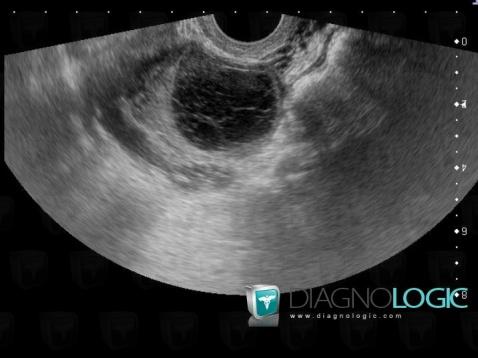 If clinical conditions and scanner availability permit, magnetic resonance imaging (MRI) is superior to CT for further characterisation of gynaecologic abnormalities, due to the excellent soft-tissue contrast, intrinsic multiplanar capabilities and lack of ionising radiation. Oblique-coronal T2-weighted (a), oblique-coronal (b) and sagittal (c) gadolinium-enhanced fat-suppressed T1-weighted images show a small-sized, unilocular fluid-containing structure in the right ovary (arrowheads), with typical homogenous T1-hypointense signal and T2-hyperintense signal, thickened walls with a crenulated enhancing rim. More common (80% of patients) compared to superficial peritoneal implants and deep pelvic endometriosis, endometrioma results from ectopic endometrial glands in the ovarian cortex that form haemorrhagic pseudocysts which become detectable by imaging. If torsion occurs in hyperstimulated ovaries, the latter may demonstrate asymmetrical augmentation, abnormal enhancement, twisted pedicle and eventually haemorrhage [50]. Cite this article. 8. DWI sequences are helpful in the characterisation of fluids: hypercellular fluids, such as pus, show restricted diffusion and therefore appear hyperintense on DWI images and hypointense on the corresponding apparent diffusion coefficient (ADC) map.
  J Ultrasound Med 22:6989, Foti PV, Attina G, Spadola S et al (2016) MR imaging of ovarian masses: classification and differential diagnosis. The normal left ovary is also seen (black arrowhead in b).
Multivariable logistic regression analysis revealed that both the presence of AB_PVP and hemoperitoneum depth were significant predictors of surgical intervention for ruptured ovarian cyst patients, with adjusted ORs of 3.773 and 1.318, respectively (Table 2). Indications include rupture, pain and haemodynamic instability. Later, a fluid-fluid level can be seen, with the dependent portion of the bloody ascites being hyperintense compared with the supernatant on T1-weighted images; on T2-weighted images, the signal intensity relationship is reversed. Compared to CT, active haemorrhage is occasionally identified on MRI as hyperintense blush on fat-suppressed T1-weighted images after gadolinium administration [8, 9]. The blood in different stages of evolution appears as iso- to hyperintense fluid on T1-weighted MR images.  Note the ipsilateral attraction of uterus (+), minimal fluid in the peritoneal cul-de sac (asterisk in a). Semin Reprod Med 28:441447, Nicholson T, Basile A (2006) Pelvic congestion syndrome, who should we treat and how? Multiplanar T2-weighted (ac) and precontrast fat-suppressed T1-weighted (df) images showed a right adnexal cyst (arrowheads) with internal fluid-fluid level and a bloody dependent component, T2-hypointense and T1-hyperintense. Right adnexal torsion in a 24-year-old woman with acute abdominal pain and leukocytosis. It is due to the invasive growth of trophoblast into the wall of the fallopian tube; the risk of rupture increases with the enlargement of the EP [40]. Alternatively, gynaecologic abnormalities (such as adnexal enlargement) may be encountered in unenhanced abdominal CT studies, such as those requested to investigate suspected urolithiasis and acute renal colic: in these situations, further investigation with contrast-enhanced CT or better MRI should be suggested after gynaecologic consultation [2, 4, 8]. One investigator reviewed the medical records of the initially selected patients and excluded the patients who were diagnosed with hemoperitoneum from causes other than corpus luteal cyst rupture (e.g., trauma, endometriosis or bleeding after ovum pick-up for in-vitro fertilization); an unruptured (incidentally found) corpus luteal cyst; or no CT scan, including 7 patients who underwent CT without a precontrast image and 2 patients whose CT images were lost due to a picture archiving and communication system (PACS) storage error. Although any functional ovarian cyst can present as a hemorrhage or rupture, the increased vascularity of the ovary in the luteal phase may increase the risk of rupture and bleeding of the corpus luteal cyst (2). Alternatively, presentation is often nonspecific with intermittent pain, low-grade fever, nausea and vomiting. Oblique coronal T2-weighted image (b) demonstrates the normal-appearing left ovary (black arrowhead). Compared to MRI, CT has poor specificity and often does not allow reliable differentiation from other complex cystic adnexal masses, either benign or malignant (Fig. However, due to fast acquisition and widespread availability on a 24/7 basis, multidetector computed tomography (CT) now represents the workhorse imaging modality in the ED and quickly provides accurate and reproducible diagnosis of most acute abdominal and pelvic complaints.
MR imaging generally demonstrates a unilocular cyst distinct from the ipsilateral ovary, hyperintense on T2-weighted images and hypointense on T1-weighted images. Our study has several limitations. To investigate patients with acute abdominal or pelvic pain, intravenous iodinated contrast agent is generally used, unless contraindicated by allergy or impaired renal function. AJR Am J Roentgenol 1989;153:747749. The amount of bleeding from a corpus luteal cyst rupture could be less than splenic bleeding, and the contrast media arrival time to cyst arteriole could be longer than the splenic artery; therefore, it is reasonable to evaluate active bleeding with a portal phase scan. Radiographics 28:16451659, Swart JE, Fishman EK (2008) Gynecologic pathology on multidetector CT: a pictorial review. In patients with torsed ovarian masses, at visual assessment, higher signal intensity of the wall of the ovarian lesion on DWI significantly correlates with haemorrhagic infarction. In the current study, the depth of hemoperitoneum was a significant CT risk factor, with an adjusted OR of 1.318. Finally, this study was a single-institute retrospective study, and a prospective multicenter study with a larger population is required to validate our results. AJR Am J Roentgenol 198:W122W131, Huang C, Hong MK, Ding DC (2017) A review of ovary torsion. At CT, the unilaterally enlarged ovary (usually greater than 5cm) is displaced from its expected site and often located on the midline, and the uterus is attracted towards the ipsilateral side by the shortened adnexal ligament. Springer Nature. Paraovarian or paratubal cysts are unilocular cystic structures located in the broad ligament, between the fallopian tube and the ovary. Additionally, sagittal T2-weighted image (c) demonstrates a predominantly low-signal haemorrhagic fluid collection (asterisk) in the pouch of Douglas. Copyright 2017 The Korean Society of Radiology. In this regard, it is important to remember the importance of post-processing images when facing a cystic mass that is hyperintense on unenhanced T1-weighted fat-suppressed sequences. Note the ovarian vessels (arrow in d), Haemorrhagic corpus luteum and functional cyst in a 15-year-old woman with pelvic pain. One patient, who had acute appendicitis and underwent removal of suspicious ruptured ovarian cyst during appendectomy, was also excluded because the decision for treatment strategy of the ruptured ovarian cyst was interrupted by the appendicitis, which required surgical treatment. Although very complete, the abovementioned protocol (Table1) lasts approximately 30min (40min including urographic acquisitions) and seems hardly applicable in an emergency setting. It is one of the most useful CT findings for the diagnosis of nutcracker syndrome, with a sensitivity of 91.7% and a specificity of 88.9% according to the literature [66, 67]. In state-of-the art multidetector scanners, CT generates almost isotropic sub-millimetre voxels that allow reconstruction of high-resolution images along arbitrary planes. Boscak AR, Shanmuganathan K, Mirvis SE, Fleiter TR, Miller LA, Sliker CW, et al. There were no significant differences in age, coitus history within 24 hours, cyst size, cyst shape (shrunken vs. maintained), presence of sentinel clot sign, presence of ring of fire sign, and attenuation/attenuation difference of the hemoperitoneum between the two treatment groups. Congested pelvic veins without AT are the hallmark of the pelvic congestion syndrome (PCS), an underdiagnosed condition that affects 9% of premenopausal women and may be exacerbated by intra-abdominal pressure. Although less prevalent than obstetric issues, acute gynaecologic disorders in non-pregnant women are not uncommon in busy emergency departments (ED). Eur J Vasc Endovasc Surg 53:886894, Hangge PT, Gupta N, Khurana A et al (2018) Degree of left renal vein compression predicts nutcracker syndrome.
First, we delineated the CT findings of ruptured corpus luteal cysts from our results. 14). Privacy Taiwan J Obstet Gynecol 2009;48:108112. The optimal cut-off value of the hemoperitoneum depth was 5.8 cm with 75.0% sensitivity and 58.6% specificity (Fig. Fertil Steril 108:886894, Jeong YY, Outwater EK, Kang HK (2000) Imaging evaluation of ovarian masses. Unilateral acute pelvic pain and tenderness leading to ED admission may result from cyst enlargement, haemorrhage or rupture. Considerable overlap exists between imaging appearances of haemorrhagic follicular and luteal cysts, and differentiation among the two entities should rely on the current phase of the menstrual cycle. After precontrast images were obtained from the diaphragmatic dome to the symphysis pubis, low-osmolar nonionic iodine contrast media (iohexol, Omnihexol 350, Korea United Pharm Inc., Seoul, Korea) was intravenously administered in a volume of 2 mL/kg using a power injector (Optivantage DH, Mallinckrodt Imaging Solutions, Hazelwood, MO, USA) at a rate of 3 mL/s. Another study on the CT findings of uncomplicated corpus luteal cysts (9), found that 6 of 10 cases (60%) showed a ring of fire sign on a CT scan. At MRI, haemoperitoneum shows variable signal intensity depending on its age (haemoglobin degradation stages), the extent of the bleeding and blood clot formation. The positive sentinel clot sign was determined as focal high-density clotted blood around the ovarian cyst on the precontrast CT scan (Fig. The cytotoxic oedema and haemorrhage that occur in ovarian torsion with haemorrhagic infarction would explain the restricted diffusion and low ADC values [53]. Borders RJ, Breiman RS, Yeh BM, Qayyum A, Coakley FV. 5). Subacute haemorrhage shows high signal intensity on fat-saturated T1-weighted images and may involve the periphery of the enlarged ovary, as T1-hyperintense rim, or the central stroma. It should also be remembered that enlarged hyperstimulated ovaries are themselves at risk for torsion [61]. At CT, the ruptured corpus luteum is seen as a shrunken ovarian cyst with discontinuity of the thickened wall, often surrounded by hyperattenuating sentinel clot. The detailed parameters were as follows: detector configuration, 16 1.25 mm; tube voltage, 120 kVp; noise index, 12.35 with automatic exposure control (smart mA, GE Healthcare, Milwaukee, WI, USA); gantry rotation period, 0.6 second; pitch factor, 1.375; table speed, 27.5 mm per rotation; reconstructed section width, 3.75 mm; and reconstructed section interval, 3.75 mm. MRI usually shows a distended dilated fallopian tube with thickened walls; the tube may demonstrate a vortex-like appearance (due to more twists) distant from the ipsilateral ovary, and the latter appears normal. described a perifollicular T2 hypointense rim correlating with perifollicular haemorrhage; the absence of this finding should be useful as a predictor of ovarian viability [51]. Bottomley C, Bourne T. Diagnosis and management of ovarian cyst accidents. Unilateral massive ovarian oedema in a 23-year-old woman with recurrent self-limiting episodes of acute pelvic pain in the last year. The ring of fire sign, which originated from the color Doppler ultrasonographic finding of a corpus luteal cyst as an increased cyst wall flow, was determined as a more prominent enhancement of the possibly involved cyst wall, with visual comparison of the myometrium as an internal reference (Fig. Emerg Radiol 25:5159, Lourenco AP, Swenson D, Tubbs RJ et al (2014) Ovarian and tubal torsion: imaging findings on US, CT, and MRI. Sagittal (a) and oblique-coronal (b) T2-weighted images show bilateral endometriomas with fluid-blood levels (arrowheads) and low signal intensity in the declivous portion of the cyst (shading sign, arrows). a, b Axial (a) and coronal (b) CT images show enlarged left ovary (arrowheads) with thickened oedematous periphery (thin arrows), containing a mixed-attenuation roundish mass with fat-attenuation foci and a calcification, corresponding to a mature cystic teratoma. df Unenhanced (d) and post-contrast (e, f) CT images show pelvic blood (asterisk) surrounding a 3-cm cystic lesion of the right ovary (arrowheads), from which contrast extravasated (arrows in e and f) indicating active bleeding. c, d Ovarian carcinoma in a premenopausal woman with predominantly cystic pattern, septations (thin arrow in d) and a solid, enhancing eccentric mural vegetation (asterisk in c). Semin Intervent Radiol 25:361368. 7). A specific CT finding is the sentinel clot sign: the presence of a hyperdense clot (>60 HU) within the adnexum, thus confirming the source of bleeding [35, 40]. Suggestive ancillary findings include multiplicity, bilateral adnexal involvement (kissing ovaries sign, Fig. The analysis of CT findings included cyst size, cyst shape, sentinel clot sign, ring of fire sign, hemoperitoneum depth, active bleeding in portal phase and attenuation of hemoperitoneum. In this early stage, ovarian stroma shows enhancement after contrast agent administration [9, 42, 45, 47,48,49,50]. In postmenopausal women, most institutions perform a preliminary unenhanced acquisition followed by optional arterial and mandatory portal venous phase CT scanning.